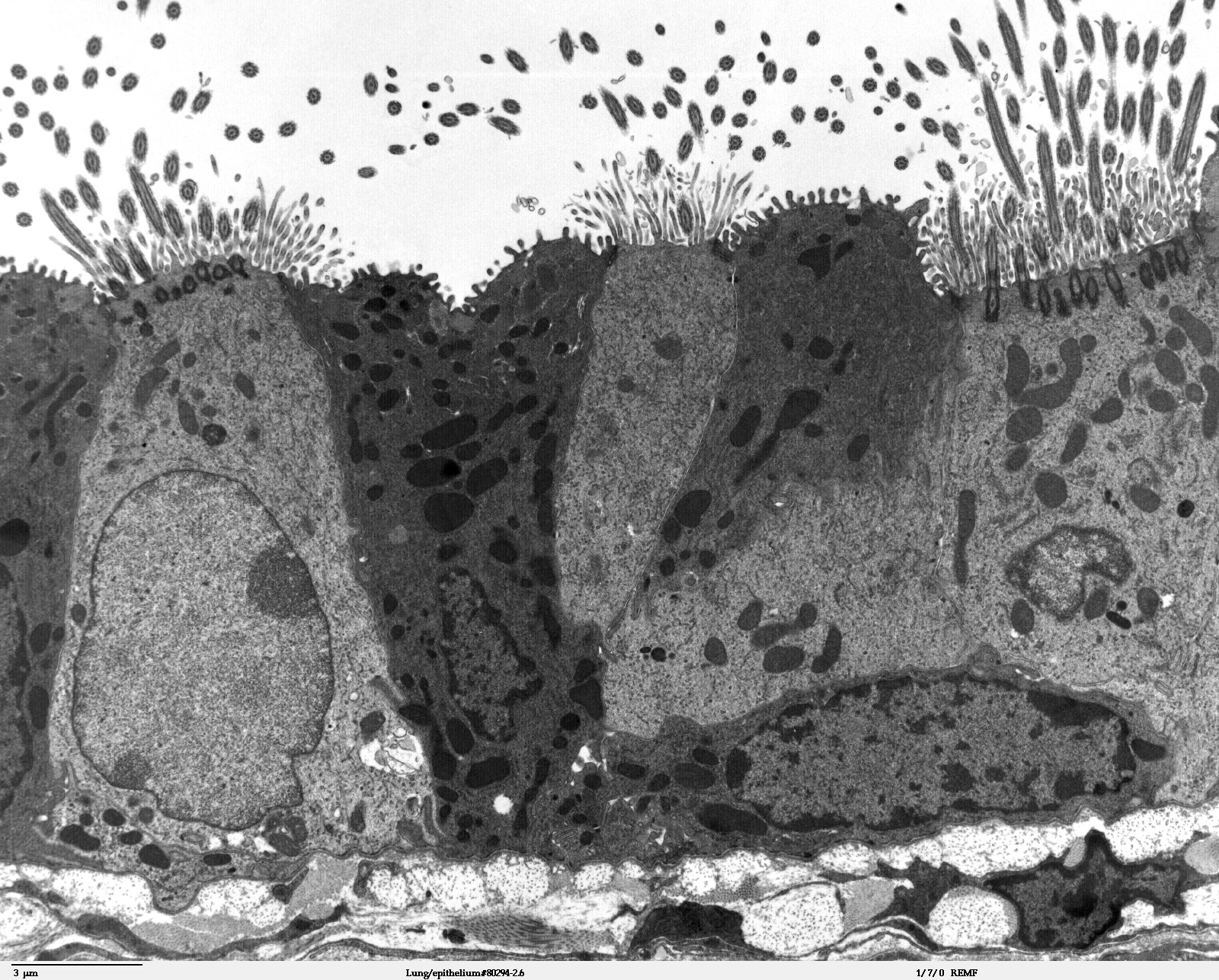
Lung epithelium 80294-2.6
Transmission electron microscope image of a thin section cut through the bronchiolar epithelium of the lung (mouse), which consists of ciliated cells and non-ciliated cells (called Clara cells). Image shows the ciliary microtubules in transverse and oblique section. In the cell apex are the basal bodies that are the anchoring sites for the ciliary axonemes. Note the difference in size and shape between the microvilli and the cilia.
JEOL 100CX TEM


Relevante Bilder


Relevante Artikel
ZelloberflächeDurch die Zellmembran wird das Innere einer Zelle vom Äußeren abgegrenzt. Die Zelloberfläche ist die Kontaktfläche, an der eine Zelle mit der Außenwelt in Kontakt tritt. Die Zelloberfläche ist genauso der Teil einer Zelle, der bestimmt, wie andere Zellen, Blutbestandteilen wie Antikörper, Komplementproteine, Hormone oder Nährstoffen mit einer gegebenen Zelle in Wechselwirkung treten. .. weiterlesen