Lower pons horizontal KB
Autor/Urheber:
Marshall Strother User:mcstrother
- Brain_stem_sagittal_section.svg: Patrick J. Lynch, medical illustrator
Attribution:
Das Bild ist mit 'Attribution Required' markiert, aber es wurden keine Informationen über die Attribution bereitgestellt. Vermutlich wurde bei Verwendung des MediaWiki-Templates für die CC-BY Lizenzen der Parameter für die Attribution weggelassen. Autoren und Urheber finden für die korrekte Verwendung der Templates hier ein Beispiel.
Shortlink:
Quelle:
Größe:
1661 x 1079 Pixel (504571 Bytes)
Beschreibung:
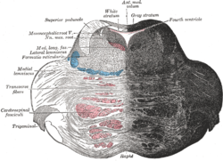
Diagram of a cross-section taken horizontally through the lower part of the pons of a human brainstem and stained with the Kluver-Barrera method. Due to the staining method, white matter (axons) appears blue and gray matter (cell bodies) appears light gray.
Notes:
- Anterior is down, posterior is up.
- The spinal tract of the trigeminal nerve can sometimes be seen surrounding the spinal nucleus of the trigeminal nerve, but it was impossible to distingusih from the inferior cerebellar peduncle on the original slide.
- The spinal lemniscus (including the spinothalamic tract) and trapezoid body also could not be distinguished but probably lie anteromedially to the superior olivary nucleus, blending in with the central tegmental tract and medial lemniscus.
- The pontine reticular formation (#9) and pontine nuclei (#22) are large, diffuse structures.
Lizenz:
Credit:
Relevante Bilder
Relevante Artikel
PonsDer Pons ist ein Abschnitt des Gehirns, der zusammen mit dem Kleinhirn zum Metencephalon (Hinterhirn) gehört. .. weiterlesen


