Onkozytom


Bei Onkozytomen (auch oxyphiles Adenom) handelt es sich um eine Gruppe gutartiger epithelialer Tumoren, die histologisch den Aufbau aus feingranulierten, großen, eosinophilen mitochondrienreichen Tumorzellen, den sogenannten Onkozyten („Hürthle-Zellen“), gemeinsam haben.
Onkozytome kommen in den Speicheldrüsen, der Niere, der Adenohypophyse und der Schilddrüse vor. Insbesondere in letzterer Lokalisation wird der Tumor nach dem Physiologen Karl Hürthle, auch als Hürthle-Zelladenom bezeichnet.
Das Onkozytom macht circa zwei Prozent der Speicheldrüsentumore aus.[1] Eine Assoziation zum Warthin-Tumor ist häufig.[1]
Diagnostik
Das Onkozytom der Niere wird gelegentlich zufällig in der Computertomographie entdeckt. Die Unterscheidung von einem malignen Tumor wie dem Nierenzellkarzinom ist in der Bildgebung oft nicht einfach, auch wenn Charakteristika wie eine zentrale Narbe oder ein sogenanntes Radspeichenphänomen beschrieben worden sind.[2]
Literatur
- Eiss et al. Renal oncocytoma: CT diagnostic criteria revisited J Radiol. 2005 Dec;86(12 Pt 1):1773-82. PMID 16333226
- Radopoulos et al. A rare case of renal oncocytoma associated with erythrocytosis: case report. BMC Urol. 2006 Sep 23;6:26. PMID 16995951
- Kosuda et al. Iodine-131 therapy for parotid oncocytoma. J Nucl Med. 1988 Jun;29(6):1126-9. PMID 3373321
Einzelnachweise
- ↑ a b W. Böcker: Pathologie : mit Zugang zum Elsevier-Portal. 5. Auflage. Urban & Fischer in Elsevier, München 2012, ISBN 978-3-437-42384-0.
- ↑ Choudhary et al.: Renal oncocytoma: CT features cannot reliably distinguish oncocytoma from other renal neoplasms. Clin Radiol. 2009;64(5):517-22. PMID 19348848
Auf dieser Seite verwendete Medien
Autor/Urheber: Die Autorenschaft wurde nicht in einer maschinell lesbaren Form angegeben. Es wird KGH als Autor angenommen (basierend auf den Rechteinhaber-Angaben)., Lizenz: CC BY-SA 3.0
Histopathological image of renal oncocytoma. Nephrectomy specimen. Hematoxylin & esoin stain.
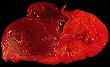
Oncocytoma of the Salivary Gland
This lesion presented as a lateral anterior neck mass. At surgery, it was found to be a soft 3.0 x 2.1 x 1.8 cm tumor of the submandibular salivary gland. The photo shows the characteristic dark color of an oncocytoma, a rare type of benign neoplasm, at the left side of the image (the normal lobulated salivary gland tissue is to the right). Excision is curative.
Since this specimen was photographed in the fresh state, it is various shades of red due to blood staining. A little formalin fixation would be expected to better emphasize the color difference between the tumor and the normal gland tissue. The photo was shot with a Minolta X-370 with 100mm Rokkor bellows lens on Kodak Elite ISO 100 daylight-balanced transparency film. I used a blue filter to compensate for the tungsten illumination.
Photograph by Ed Uthman, MD. Public domain. Posted 19 May 00Autor/Urheber: Hellerhoff, Lizenz: CC BY-SA 3.0
Onkozytom der rechten Niere (im Bild links) in der Computertomographie. Typisches Radspeichenphänomen. die meisten Onkozytome sind jedoch ct-morphologisch nicht von anderen Nierentumoren, insbesondere vom klarzelligen Nierenzellkarzinom zu unterscheiden.


