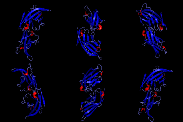
B7-2
| T-lymphocyte activation antigen CD86 | ||
|---|---|---|
 | ||
| lösliche Variante von B7-2 | ||
| Andere Namen | Activation B7-2 antigen, B70, BU63, CTLA-4 counter-receptor B7.2, FUN-1, CD86 | |
| Eigenschaften des menschlichen Proteins | ||
| Masse/Länge Primärstruktur | 329 Aminosäuren, 37.682 Da | |
| Bezeichner | ||
| Externe IDs | ||
| Orthologe (Mensch) | ||
| Entrez | 942 | |
| Ensembl | ENSG00000114013 | |
| UniProt | P42081 | |
| Refseq (mRNA) | NM_001206924.1 | |
| Refseq (Protein) | NP_001193853.1 | |
| PubMed-Suche | 942 | |
B7-2, in der Literatur häufig auch als CD86 bezeichnet, ist ein Oberflächenprotein aus der Immunglobulin-Superfamilie. Als Kostimulator ist es an der Aktivierung von T-Zellen im Zuge der Immunantwort beteiligt.[1]
Eigenschaften
B7-2 wird von aktivierten B-Zellen und Monozyten exprimiert und bildet Homodimere.[2] Die Aktivierung über einen Kostimulator liefert das notwendige zweite Aktivierungssignal für T-Zellen. B7-2 bindet an CD28 (T-Zell-aktivierend) oder CTLA-4 (T-Zell-inaktivierend). Die Isoform 2 von B7-2 hemmt die Bindung der Isoform 1 aneinander und hemmt dadurch die Aktivierung. B7-2 ist glykosyliert und besitzt eine ähnliche Funktion wie B7-1.
B7-2 ist der zelluläre Rezeptor der Adenoviren der Gruppe B.
Weblinks
Einzelnachweise
- ↑ S. Bhatia, M. Edidin, S. C. Almo, S. G. Nathenson: B7-1 and B7-2: similar costimulatory ligands with different biochemical, oligomeric and signaling properties. In: Immunology letters. Band 104, Nummer 1–2, April 2006, S. 70–75, doi:10.1016/j.imlet.2005.11.019, PMID 16413062.
- ↑ Charles Janeway: The production of armed effector T cells. In: Immunobiology (2001). 3. Februar 2017, abgerufen am 17. Mai 2017 (englisch).
Auf dieser Seite verwendete Medien
This is the crystal structure of the soluble form of CD86. It is a necessary costimulating molecule needed to activate T cells for priming of an immune response. It is known to bind CD28 and is similar in function to CD80. The original Protein Data Bank (pdb) file was published by Zhang et al (2003). I animated the pdb using PyMol. For the original pdb, see: Zhang, X., Schwartz, J.D., Almo, S.C., Nathenson, S.G. Crystal Structure of the Receptor-Binding Domain of Human B7-2: Insights into Organization and Signaling Proc.Natl.Acad.Sci.USA v100 pp.2586-2591 , 2003
